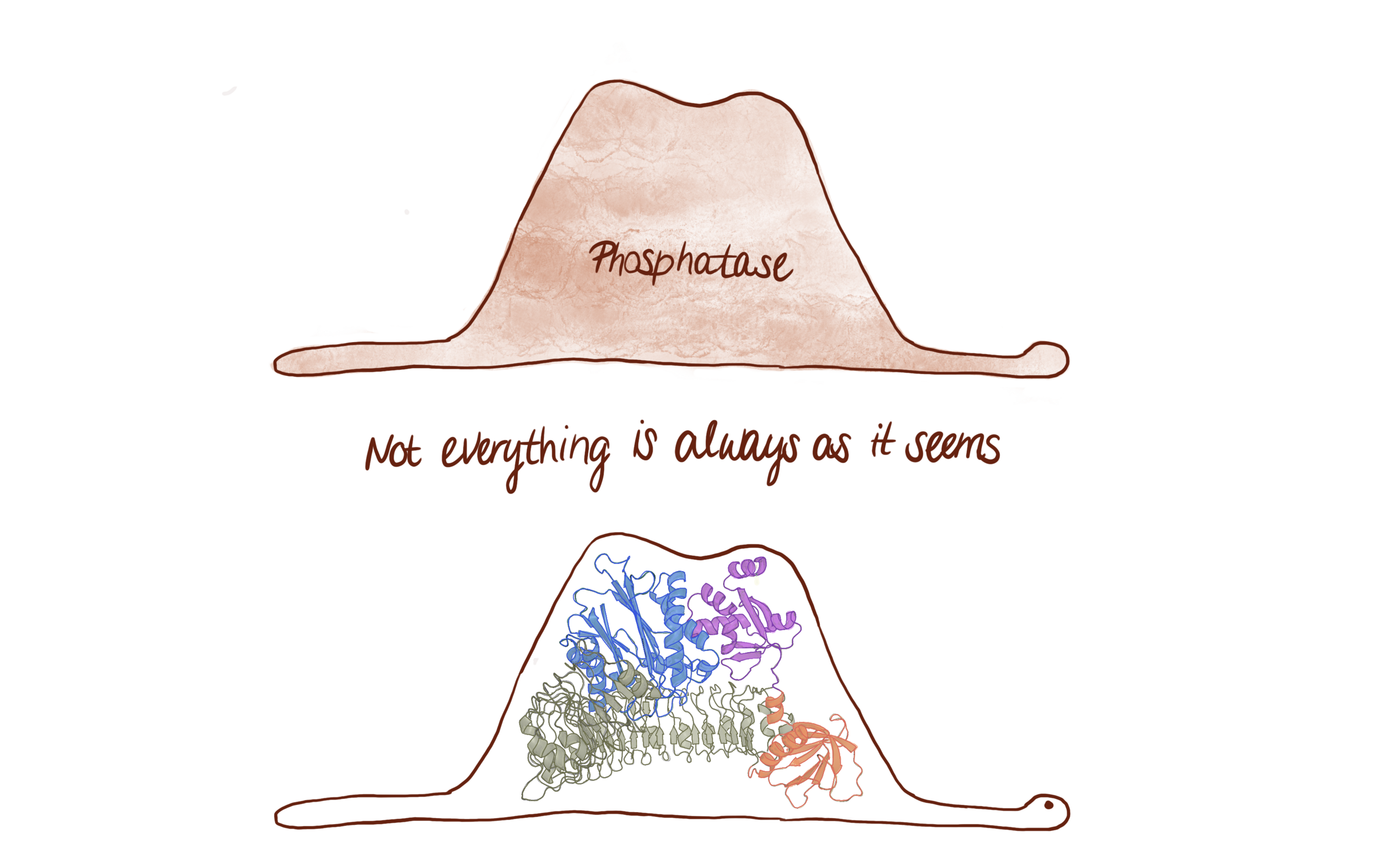
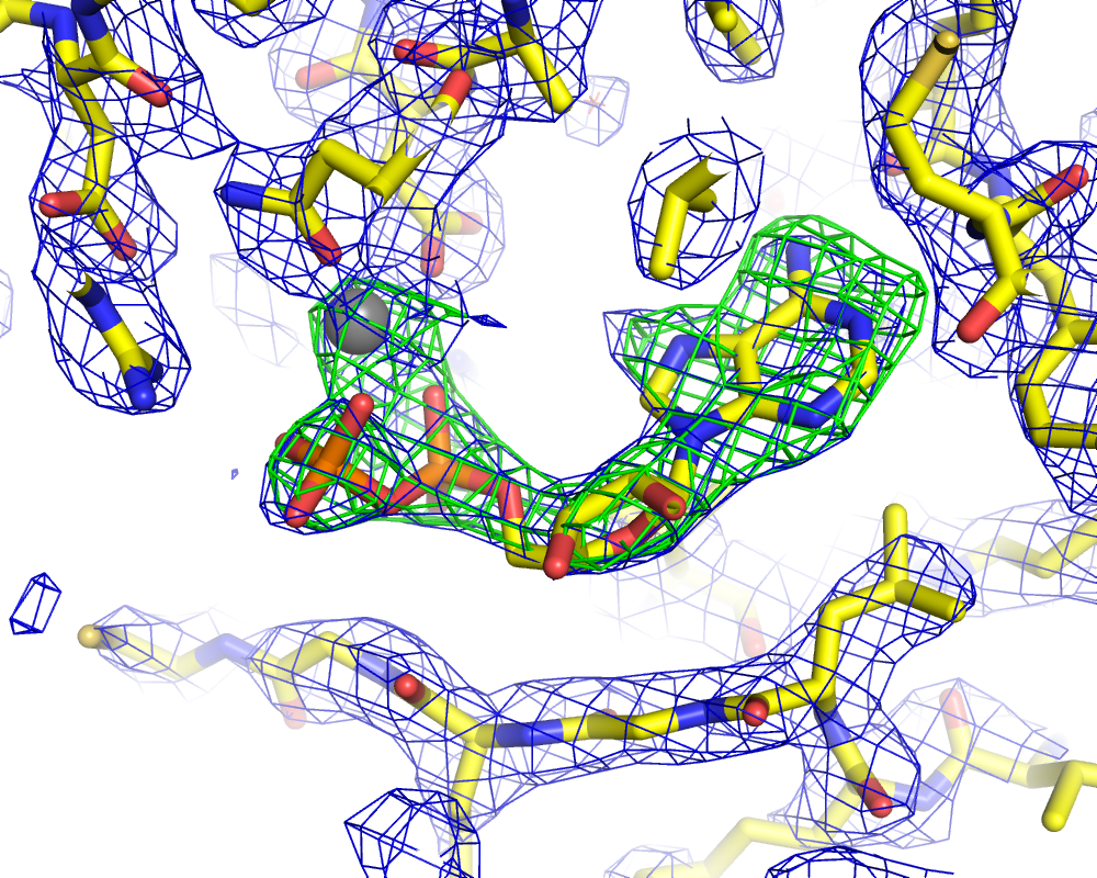
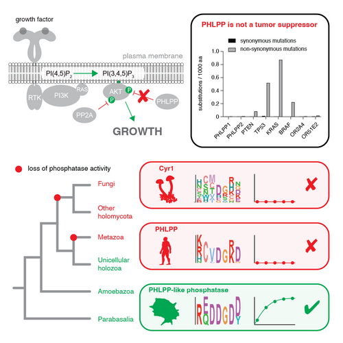
On this page
Physiological processes are driven by the coordinated action of organs, tissues and cells. Coordination of these activities allows organisms to respond appropriately to their changing environment. Within cells themselves, important metabolic processes are often compartmentalised by membranes, which necessitates the flow of information between compartments. How is this information integrated into the appropriate downstream response? We try to understand the basic principles that govern the flow of information in cells with particular emphasis on the role of lipid second messengers. By understanding the mechanisms by which these signals are transduced and propagated, we can better answer why and how things go wrong in disease.
We study how lipids regulate the activity of signaling enzymes, including both protein kinases and phosphatases. We use biochemical and cell biological assays supported by biophysical and structural techniques to deduce how, where, and when the signal is transduced. Our work is underpinned by quantitative biochemistry performed with precisely defined macromolecules. The insights we derive have the potential to rationalize disease pathogenesis at the molecular and atomic levels. For example, in the pro-growth and survival kinase Akt, we have characterized the mechanism by which a mutation associated with cancer and overgrowth disorders leads to kinase activity that is both spatially and temporally de-regulated. Determining how specific mutations drive the development of disease is essential for effective therapeutic intervention.
Thomas studied Biochemistry at the University of Bristol, U.K. before obtaining a PhD in Structural Biology at the MRC Laboratory of Molecular Biology in Cambridge. Thomas moved to the USA in 2005 as an EMBO postdoctoral fellow at the National Institutes of Health in Bethesda, MD and started his own lab as an independent investigator at the Max Perutz Labs in 2012.
The regulation of protein kinase activity by activation loop phosphorylation is a well-characterized mechanism. Protein kinases that auto-phosphorylate usually do so in trans: dimerization results in reciprocal phosphorylation of the activation loop by the other protomer. However, we have discovered that Protein Kinase D (PKD) has evolved the inverse solution to the same problem. PKD is an inactive dimer that is activated by kinase domain dissociation followed by cis-autophosphorylation (Reinhardt et al, PNAS, 2023).
The myotonic dystrophy kinases of the DMPK family are conserved regulators of actomyosin contractility. The four prototypical DMPK members are obligate homodimers with a conserved architecture of N-terminal kinase domains, a central coiled-coil, and C-terminal membrane binding domains. Remarkably, the length of the coiled-coil domain of each family member varies between 10 and 145 nm, but is astonishingly well conserved. We present a model in which the activity of these kinases is regulated by their spatial positioning with respect to the membrane (Truebestein et al, Structure, 2023).
Phosphoinositide-dependent kinase 1 (PDK1) is an essential protein kinase that controls cellular and organismal growth. We have elucidated the mechanism by which PDK1 is activated by phosphatidylinositol-3,4,5-trisphosphate (PIP3) at the plasma membrane in response to growth factor-mediated activation of cell surface receptors. Our work reveals previously unknown mechanisms of autoinhibition and trans-autoactivation, with opportunities for the development of novel therapeutics in the treatment of cancer (Levina et al, Nature Communications, 2022).
© Illustration by Dorotea Fracchiolla (Art&Science)
The lipid second messengers PI(3,4,5)P3 and PI(3,4)P2 have long been known to activate Akt by recruiting it to membranes, where it is activated by phosphorylation. We have determined the structure of autoinhibited Akt1, revealing the molecular basis of a disease-causing mutation in the PH domain which gives rise to Proteus Syndrome and some cancers. Phosphorylation is insufficient to fully activate Akt1. Our work implies that Akt signaling is restricted to discrete membrane locations in the cell (Truebestein et al, PNAS, 2021).
Serum- and glucocorticoid-regulated kinase 3 (Sgk3) is activated by the phospholipid phosphatidylinsoitol 3-phosphate (PI3P) in the PI3 kinase pathway. Up-regulation of Sgk3 activity has increasingly been implicated in cancers that exhibit paradoxically low Akt activity. We have discovered that Sgk3 is dependent on PI3P for its activity, which restricts its activity to PI3P-rich endosomes in cells. We have biochemically reconstituted Sgk3 activation downstream of the class III PI 3-kinase Vps34 in vitro (Pokorny et al, Journal of Biological Chemistry, 2021).
Protein Kinase D (PKD) is an essential ser/thr kinase involved in vesicular transport of cargo from the trans-Golgi network to the plasma membrane. We have discovered a novel, ubiquitin-like domain in PKD that mediates its dimerization on diacylglycerol-containing membranes, thereby driving trans-autophosphorylation and activation of its kinase domain (Elsner et al, Journal of Biological Chemistry, 2019).
The protein kinase Akt integrates signals from membrane-embedded lipid second messengers and signals in the form of phosphorylation by upstream protein kinases. We have shown that one without the other is insufficient for Akt activation and that membrane dissociation triggers rapid dephosphorylation and inactivation of Akt (Lučić et al, PNAS, 2018).
We recently demonstrated that the activity of a protein kinase called Akt, which promotes cell growth and proliferation, is strictly governed by the engagement of two specific lipids in cell membranes (Ebner & Lučić et al, Molecular Cell, 2017). Bypassing this requirement leads to uncontrolled growth and cancer.
We have discovered an allosteric switch in the Src and Tec kinases that converts them into active enzymes by promoting the exchange of ADP for ATP (von Raußendorf et al, Scientific Reports, 2017). The switch depends on recognition of a polyproline motif in the tail of activated receptors at the plasma membrane.
Rho-associated coiled-coil kinase (ROCK) is a key signal transducer in regulating the cytoskeleton. Substrate engagement is controlled by the precise positioning of its kinase domains, which is achieved via a long coiled-coil domain that bridges its membrane binding domains to its kinase domains (Truebestein et al., Nature Communications, 2016).
Membranes are sites of intense signaling activity in eukaryotic cells. Essential processes such as autophagy, cytokinesis, exo- and endo- cytosis, and cytoskeletal remodeling depend on signal propagation at cellular membranes. Dysregulation of signal transduction at these sites is the cause of a number of hereditary and non-hereditary diseases, including Coffin-Lowry syndrome, spinocerebellar ataxia, myotonic dystrophy, and various cancers. Over 500 kinases and 130 phosphatases regulate signal transduction by phosphorylating or dephosphorylating their target proteins. Given that the chemistry of phosphoryl transfer is conserved, there is a clear need for compartmentalization of what are essentially the same chemical reactions.
One of the most important consequences of the activation of cell surface receptors is the generation of small molecule second messengers. In addition to the freely diffusible second messengers such as cAMP and inositol-1,4,5-triphosphate (IP3), a number of cellular second messengers are lipids. Despite being of fundamental importance to the exquisite spatial and temporal regulation of many cellular processes, the molecular mechanisms of lipid-mediated signal transduction are not well understood. Our goal is to understand how lipid second messengers can turn on signaling pathways at the membrane. To achieve this, we are using a spectrum of biophysical (including X-ray crystallography), biochemical, and cell biological techniques. Fundamentally, we aim to describe signal transduction processes with deep mechanistic insight. By combining structure with quantitative in vitro biochemistry on rigorously validated samples, we can probe the structure-function relationship in absolute terms.
Many of the lipid responsive human protein kinases belong to the AGC family of kinases, of which paradigmatic lipid-activated kinases are Akt, PDK1 and Sgk3. In 2017 we demonstrated the allosteric activation of Akt by the lipid second messengers PI(3,4,5)P3 and PI(3,4)P2 for the first time. A mutation in the kinase domain associated with cancer and overgrowth disorders of the brain causes Akt hyperactivation by relieving autoinhibition (Ebner and Lučić et al., Molecular Cell 2017). We have since described the mechanism of activation in more detail (Truebestein et al., PNAS 2021) and characterized the allosteric coupling between PH domain-mediated autoinhibition and the inactivation of Akt by dephosphorylation (Lučić et al., PNAS 2018). Conceptually, Akt can be thought of as an AND gate that integrates multiple signals to drive the biological response.
Similarly, Sgk3 employs similar mechanisms of autoinhibition and allosteric activation to drive signaling downstream of the lipid second messenger PI3P. Like Akt, Sgk3 exhibits an inactive conformation in the absence of PI3P in which the PI3P-binding pocket is sequestered in an intramolecular interface between the regulatory and kinase domains (Pokorny et al, Journal of Biological Chemistry 2021). Akt and Sgk3 activities are thereby restricted, exclusively, to those membranes in the cell where they can be activated by their cognate lipids.
Protein kinases are often activated by autophosphorylation of a key residue in their activation loop that drives catalytic activity. Mechanistically, this reaction may occur in trans or in cis. In 2019, we determined the structure of a novel ubiquitin-like domain in Protein Kinase D (PKD) which mediates its dimerization (Elsner et al., Journal of Biological Chemistry 2019). PKD, however, is maintained in an inactive conformation by dimerization and relief of this trans-autoinhibition leads to activation loop autophosphorylation in cis (Reinhardt et al., PNAS 2023). PKD is an essential mammalian kinase involved in trafficking of cargo destined for secretion from the trans-Golgi network to the plasma membrane.
In a parallel study on phosphoinositide-dependent kinase 1 (PDK1), which is a master regulator of growth factor signaling, we have determined the mechanism of its activation by PIP3-elicited dimerization and trans-autophosphorylation (Levina et al., Nature Communications 2022). These studies showcase how nature has employed common strategies, such as dimerization and activation loop phosphorylation, in different ways to control signal transduction.
In work on the Tec and Src family tyrosine kinases, we have revealed a previously unknown switch in nucleotide binding that drives kinase activation (von Raußendorf et al., Sci Rep 2017). Here nature utilizes ADP, the product of the kinase reaction, to stabilize and maintain the inactive conformation. Upon triggering of the conformational changes required for kinase activation, these kinases lose their strict preference for ADP, which is then rapidly exchanged for ATP in the cell. The binding of ADP in the inactive conformation serves to insulate the kinase from promiscuous activity, while activation drives the exchange of ADP for the ATP required for substrate phosphorylation.
We are also interested in how signal transduction pathways are organized. Scaffolding of signaling proteins in the same pathway enhances specificity, promotes signal amplification by reducing noise, and, ultimately, improves signal propagation through the pathway. Membranes act as the scaffolds for many signaling reactions, including those involved in regulation of the actin cytoskeleton (Truebestein et al., Nature Communications 2016; Truebestein et al., Structure 2023). Our studies are aimed at understanding how diverse signals are integrated, how substrate specificity is encoded not just at the kinase level, and the influence of the membrane environment on multi-component signaling hubs. This is an exciting area of research with frontiers in ageing, cancer, metabolic diseases such as diabetes, and obesity.
Ronja defended her PhD thesis with an outstanding explanation of kinase activation by autophosphorylation that was understandable to a non-expert audience. Ronja's work on Protein Kinase D (PKD) has revealed an unexpected mechanism of kinase regulation by trans-autoinhibition, which is relieved by a combination of membrane binding and cis-autophosphorylation. Ronja's work has major implications for the regulation of other protein kinases that are widely believed to be activated by trans-autophosphorylation.
In this Review Article, just published in eLife, we examine the mechanistic basis for protein kinase activation by autophosphorylation. We critically evaluate the evidence for both cis (intramolecular) and trans (intermolecular) mechanisms, summarise the advantages and pitfalls of common experimental approaches, and encourage more quantitative and specific reporting.
PhD students of the Laboratory of Molecular Biology (LMB), Cambridge and the Vienna BioCenter (VBC) have teamed up to organize this year's LMB-VBC Graduate Life Sciences Symposium. This international symposium showcases cutting-edge research in the fields of structural biology, neuroscience, immunology, nucleic acids, and cell biology. Ronja Reinhardt (Leonard lab) will present her recently published work on Protein Kinase D regulation.
In this Review Article, just published in Developmental Cell, Thomas Leonard, Martin Loose and Sascha Martens discuss membrane-localized reactions with a particular focus on insights derived from reconstituted systems. The authors explain how the interplay of regulatory proteins results in the self-organization of cellular factors, their condensation, assembly, and activity.
The Leonard Lab has been awarded a grant by the Austrian Agency for Education and Internationalisation (OeAD) to foster cooperation between researchers in Austria and Bulgaria. The grant will support scientific exchange between the Max Perutz Labs and the University of Sofia. The Leonard lab will collaborate with the lab of Prof. Todor Dudev, an expert in the chemistry of metalloproteins, to investigate the biochemistry of a key signal transducer and reported tumor suppressor.
The Leonard Lab has been awarded a new grant by the Austrian Science Fund. If you are interested in understanding how signals are encoded, decoded, and transduced into approriate downstream responses in cells, please get in touch. We have projects at Master, PhD and Postdoc level just waiting for the right people!
In recent years, Akt has emerged as a clinical target in the treatment of various cancers and overgrowth disorders. Two types of inhibitors have been developed to specifically target Akt: ATP-competitive inhibitors and allosteric inhibitors, which recognize a unique interface between its regulatory, PIP3-binding PH domain, and its kinase domain. Using HDX-MS, John Burke's lab (University of Victoria, British Columbia) now reveals the conformational changes induced by inhibitor binding, the results of which may inform the future development of Akt-targed therapeutics (Shaw et al, Structure, 2023).
The PI is perfectly normal - honestly. 2 years after his students bought him a bungee jump for becoming a tenured Professor at the Max Perutz Labs, Thomas thanked them all and stepped off Jauntalbrücke in Carinthia, Austria for a 96 m plunge towards the river below. Sadly, none of his students could be convinced to jump with him. Check out the video to see what students can do for their PI!
Structure and regulation of the myotonic dystrophy kinase-related Cdc42-binding kinase.
Truebestein, Linda; Antonioli, Sumire; Waltenberger, Elisabeth; Gehin, Charlotte; Gavin, Anne-Claude; Leonard, Thomas A
PKD autoinhibition in trans regulates activation loop autophosphorylation in cis.
Reinhardt, Ronja; Hirzel, Kai; Link, Gisela; Eisler, Stephan A; Hägele, Tanja; Parson, Matthew A H; Burke, John E; Hausser, Angelika; Leonard, Thomas A
Activation of the essential kinase PDK1 by phosphoinositide-driven trans-autophosphorylation.
Levina, Aleksandra; Fleming, Kaelin D; Burke, John E; Leonard, Thomas A
Structure of autoinhibited Akt1 reveals mechanism of PIP3-mediated activation.
Truebestein, Linda; Hornegger, Harald; Anrather, Dorothea; Hartl, Markus; Fleming, Kaelin D; Stariha, Jordan T B; Pardon, Els; Steyaert, Jan; Burke, John E; Leonard, Thomas A
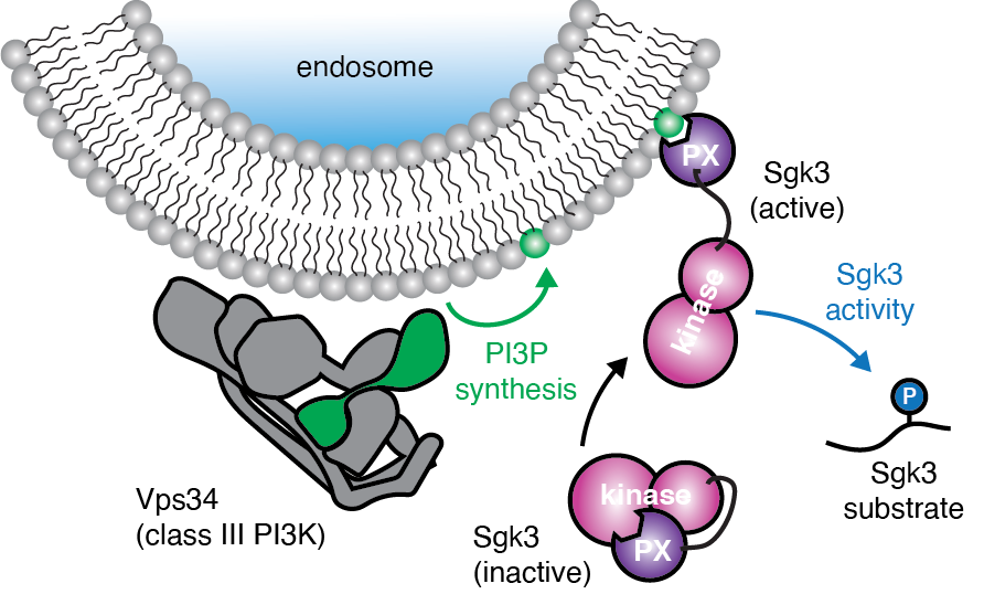
In vitro reconstitution of Sgk3 activation by phosphatidylinositol 3-phosphate.
Pokorny, Daniel; Truebestein, Linda; Fleming, Kaelin D; Burke, John E; Leonard, Thomas A
A ubiquitin-like domain controls Protein Kinase D dimerization and activation by trans-autophosphorylation.
Elsner, Daniel J; Siess, Katharina M; Gossenreiter, Thomas; Hartl, Markus; Leonard, Thomas A
Conformational sampling of membranes by Akt controls its activation and inactivation.
Lučić, Iva; Rathinaswamy, Manoj K; Truebestein, Linda; Hamelin, David J; Burke, John E; Leonard, Thomas A
PI(3,4,5)P3 Engagement Restricts Akt Activity to Cellular Membranes
Michael Ebner, Iva Lučić, Thomas A. Leonard, and Ivan Yudushkin
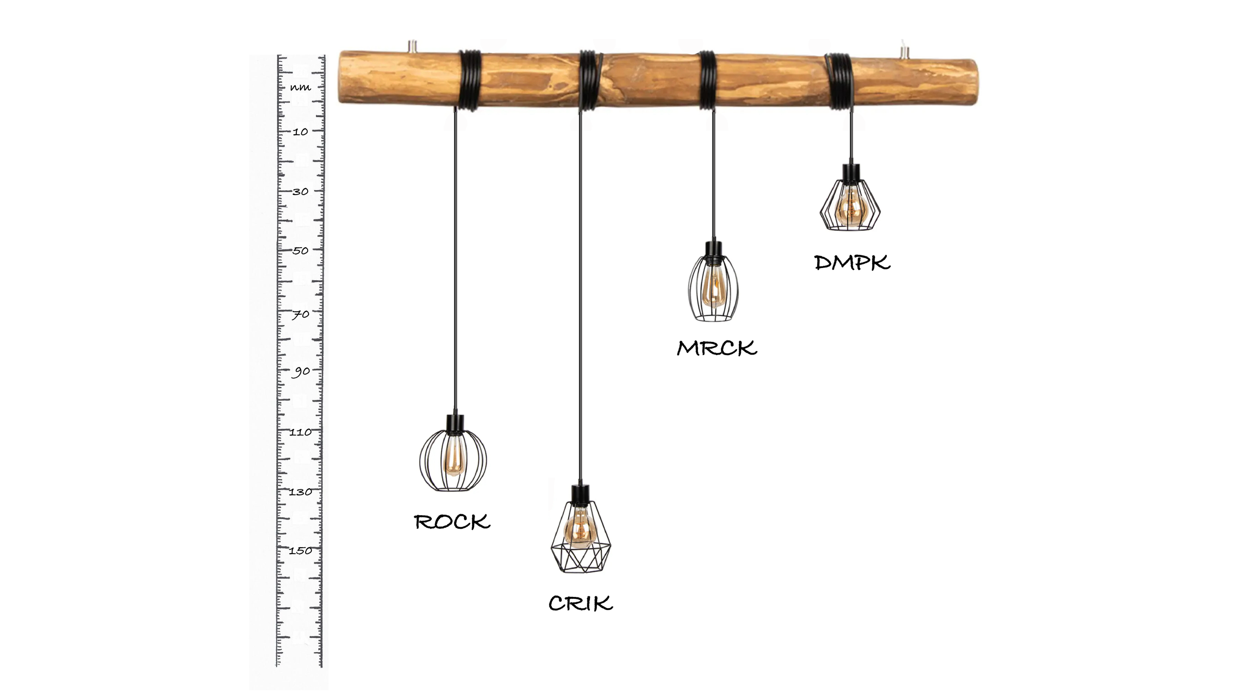
A molecular ruler regulates cytoskeletal remodelling by the Rho kinases
Truebestein, L., Elsner, D.J., Fuchs, E., Leonard, T.A.
Project title: 'Phosphoinositide-dependent kinase 1: master growth regulator' (P 36212)
Project title: 'PI3K signaling- navigating upstream and downstream of Akt' (P 33066)
Project title: 'Structure, function and regulation of Protein Kinase D' (P 30584)
The Leonard Group is a member of the special doctoral program 'Signaling Mechanisms in Cellular Homeostasis (W1261)', reviewed and funded by the Austrian Science Fund (renewed for second funding period, 2021-2025).
Project title: 'Microtubule-associated serine/threonine kinases in health and disease' (P 36724)
Project title: 'PHLPP – elucidating the mechanism of action of a tumor suppressor phosphatase' (BG 05/2023)
Phage therapy for treating bacterial infections: a double-edged sword
Suckers and segments of the octopus arm
Using the house mouse radiation to study the rapid evolution of genes and genetic processes
CRISPR jumps ahead: mechanistic insights into CRISPR-associated transposons
SLiMs and SHelMs: Decoding how short linear and helical motifs direct PPP specificity to direct signaling
Title to be announced
Enigmatic evolutionary origin and multipotency of the neural crest cells - major drivers of vertebrate evolution
Visualising mitotic chromosomes and nuclear dynamics by correlative light and electron microscopy
Engineered nanocarriers for imaging of small proteins by CryoEM
Bacterial cell envelope homeostasis at the (post)transcriptional level
Title to be announced
Hydrologic extremes alter mechanisms and pathways of carbon export from mountainous floodplain soils
Dissecting post-transcriptional gene expression regulation in humans and viruses
Polyploidy and rediploidisation in stressful times
Prdm9 control of meiotic synapsis of homologs in intersubspecific hybrids
Title to be announced
RNA virus from museum specimens
Programmed DNA double-strand breaks during meiosis: Mechanism and evolution
Title to be announced