On this page
test1 Test2
Test3
In any micrograph, the number of detected probe particles is fundamentally limited, either due to finite acquisition times or probe-induced sample damage. How can we optimize the sensitivity of a microscope and maximize the information that can be extracted from each detected probe particle?
We employ cavity enhancement, quantum enhancement, and wave-front shaping techniques to optimize the sensitivity of interferometric imaging techniques. We do so both in electron and light microscopy.
Thomas Juffmann did his PhD on molecular quantum optics at the University of Vienna. He then moved to Stanford to work on quantum enhanced imaging with electrons and photons. After that, he joined the ENS Paris as an interdisciplinary HFSP fellow working on adaptive optics. In 2018, he started the Quantum Imaging and Biophysics group in a joint venture between the Faculty of Physics and the Max Perutz Labs.
Passing probe particles through a sample multiple times can increase the signal to noise per detected particle. We demonstrated full field cavity enhanced microscopy with light [1], and are now extending this approach to iScat measurments. We also showed theoretically [2], that the same principle could enable cryogenic electron microscopy at unprecedented low damage levels. The first multi-pass TEM is now in the making [3].
[1] Multi-pass microscopy; T. Juffmann, B. B. Klopfer, T. L.I. Frankort, P. Haslinger & M. A. Kasevich; Nature Communications, Vol. 7, 12858 (2016)
[2] Multi-pass transmission electron microscopy; T. Juffmann, S. A. Koppell, B. B. Klopfer, C. Ophus, R. M. Glaeser & M. A. Kasevich; Scientific Reports, Vol. 7, 1699 (2017)
[3] Design for a 10 keV multi-pass transmission electron microscope; S. A. Koppell, M. Mankos, A. J. Bowman, Y. Israel, T. Juffmann, B. B. Klopfer, M. A. Kasevich; Ultramicroscopy, Vol. 207, 112834 (2019)
Phase microscopy is based on interfering a signal wave with a reference wave. Depending on local, sample induced phase-shifts, the sensitivity of the most common phase microscopy techniques can be far from ideal. We could show this deriving the Cramer Rao bounds in phase imaging for any linear optical system [4]. Local Wave-front shaping for Phase Imaging (Lowphi) can then be used to adaptively optimize your microscope to a specific sample [5].
[4] Fundamental bounds on the precision of classical phase microscopes; D. Bouchet, J. Dong, D. Maestre, and T. Juffmann; arXiv:2011.04799 (2020)
[5] Local Optimization of Wave-fronts for high sensitivity Phase Imaging; T. Juffmann, A. de los Rios Sommer, S. Gigan; Optics Communications, Vol. 454, 124484 (2020)
Pulsed laser deposition assisted epitaxial growth of cesium telluride photocathodes for high brightness electron sources
K.P. Mondal, M. Gaowei, E. Echeverria, K. Evans-Lutterodt, J. Jordan-Sweet, T. Juffmann, S. Karkare, J. Maxson, SJ van der Molen, C. Pennington, P. Saha, J. Smedley, WG Stam, R. M. Tromp et al.
A structural analysis of ordered Cs3Sb films grown on single crystal graphene and silicon carbide substrates
C. A. Pennington, M. Gaowei, E. M. Echeverria, K. Evans-Lutterodt, A. Galdi, T. Juffmann, S. Karkare, J. Maxson, S. J. van der Molen, P. Saha, J. Smedley, W. G. Stam, R. M. Tromp
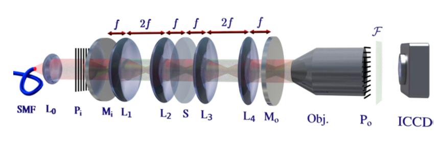
Unified Simulation Platform for Interference Microscopy
F. Hitzelhammer, A. Dostálová, I. Zykov, B. Platzer, C. Conrad-Billroth, T. Juffmann, U. Hohenester
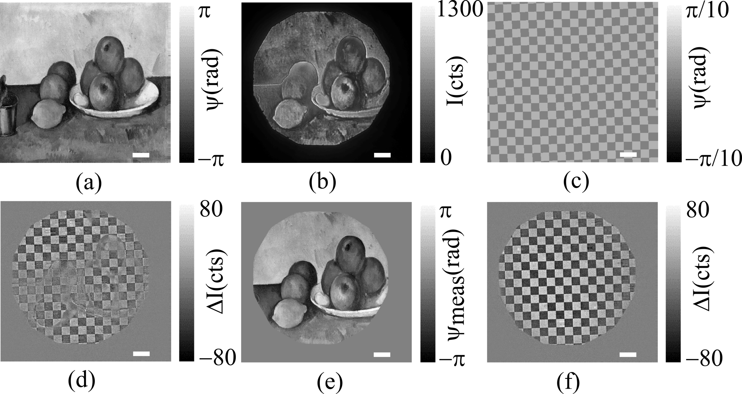
Growth of ultra-flat ultra-thin alkali antimonide photocathode films
G. WG Stam, M. Gaowei, EM. Echeverria, K. Evans-Lutterodt, J. Jordan-Sweet, T. Juffmann, S. Karkare, J. Maxson, SJ van der Molen, C. Pennington, P. Saha, J. Smedley, RM Tromp
Quantum Limits of Position and Polarizability Estimation in the Optical Near Field
L. Kienesberger, T. Juffmann, S. Nimmrichter